Magnetic micromanipulators for probing local rheological properties of scaffolds and vital 3D tissue constructs
Lead supervisor: U. Schnakenberg, Co-supervisor: S. Uhlig RWTH Aachen, Institute of Materials in Electrical Engineering 1 (IWE1)
Hypothesis: Magnetic micromanipulation techniques facilitate mapping of viscoelastic heterogeneities at high spatial resolution in vital tissues.
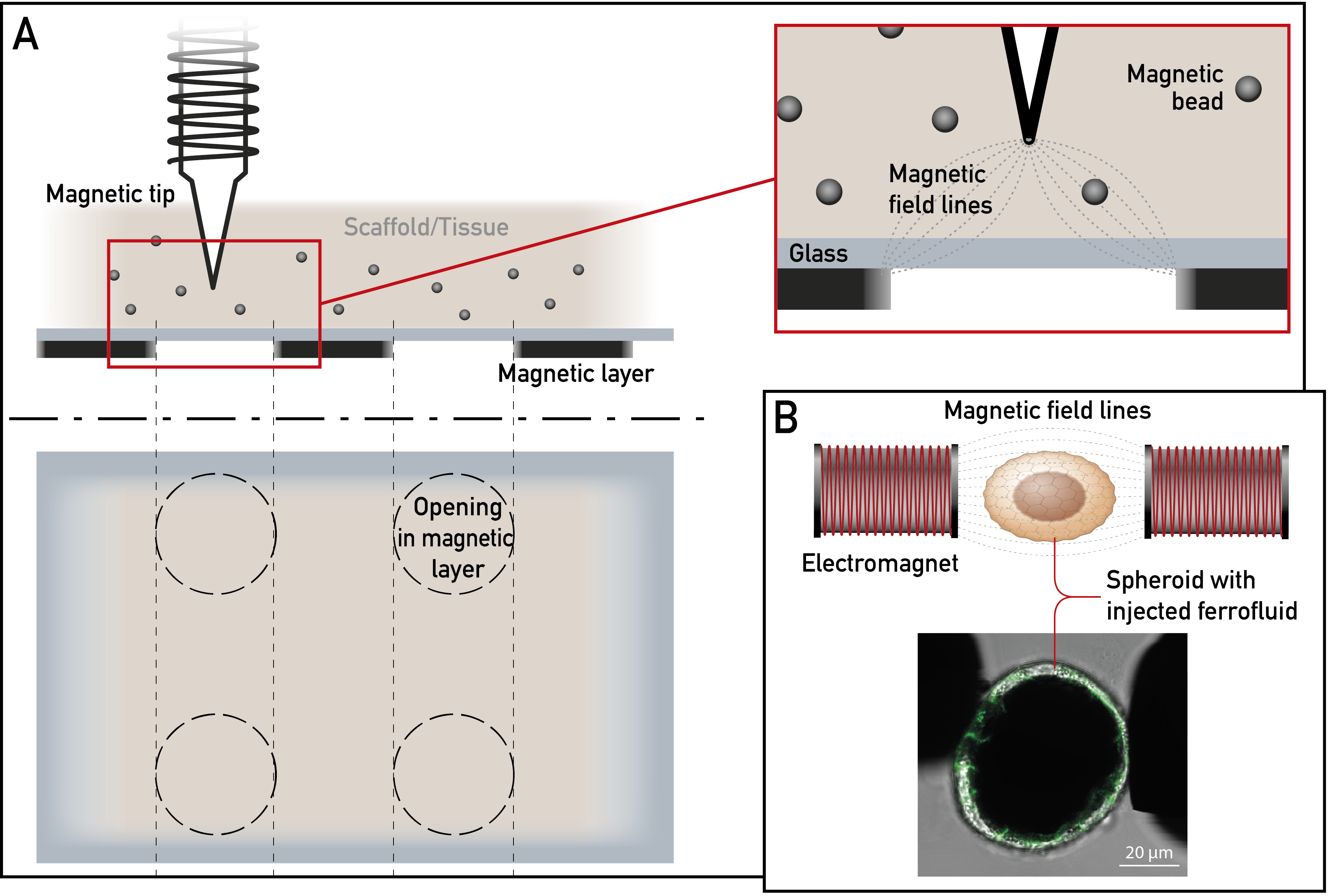
Illustration of the two major experimental approaches. (A) Optimized pencil-type magnetic tweezers for insertion in material of interest. Top left: Cross section. Top right: The magnetic field lines from the magnetic tip to the edge of the opening in the magnetic layer at the bottom of the glass slide form a well-defined magnetic field gradient. Bottom: Top view. Dotted circles indicate the circular openings in the magnetic layer below the material of interest. (B) Setup for characterization of rheological properties of epithelial spheroids filled with ferrofluid. Forces on the ferrofluid are applied with an electromagnet in a hoerseshoe configuration. Drawings not to scale. A preliminary experiment is shown presenting the deformation response of a ferrofluid-filled MDCK-derived cyst, cells transfected with cadherin-GFP (experiment carried out in collaboration with E. Noetzel-Reiss [A2] and V. Buck [A3]).
Key preliminary results of project D1. (A) Aligned nerve cell extensions from a dorsal root ganglion in a PEG-based Anisogel (unpublished). (B) Aligned primary nerve cells in a fibrin-based Anisogelfrom 39. (C) Fibroblast adhering to RGD-modified microgel from74.
Background: The mechanical properties of tissues and cells and their microenvironment are highly heterogenous and subject to changes during development and in response to various stresses. In situ and in vivo measurements of mechanical properties are therefore urgently required. Biocompatible magnetic particles and ferrofluids have shown great promise towards achieving this goal. Pencil-type magnetic tweezers are commonly used for manipulating magnetic beads101,102. A major advantage in comparison to optical tweezers is that high forces can be generated. We have used them to move ingested superparamagnetic beads to measure cytoplasmic viscoelasticity in keratinocytes (with MOCA and FZJ)60,69. To reduce the labor-intensive and challenging hands-on time of single cell measurements, we are currently developing a novel type of planar magnetic tweezers setup on glass substrates using electroplated nickel-iron layers. Deforming ferrofluid droplets with homogeneous magnetic fields is a recent ground-breaking advancement in the characterization of rheological properties of vital micro-environments103. Ferrofluids are made of magnetic nanoparticles that form highly deformable droplets when injected into living tissue. The magnetic moments of these particles orient along applied magnetic fields leading to interacting dipole moments and droplet deformation to the shape of a prolate spheroid, whose length is determined by the balance of magnetic stress and surface tension104,105. In contrast to solid microbeads, the liquid ferrofluid droplets produce less damage in vital micro-environments. Aims: The goal is to further develop tools to enable local viscoelasticity measurements at the highest possible spatial resolution in living epithelial cell assemblies. The new tools are tested in MEƎT's different culture systems and substrates. Approach: We pursue two strategies: The pencil-type magnetic tweezers setup, which is designed to apply forces on single cells and cells grown in monolayers, are adapted for use in complex 3D tissue assemblies by (i) reduction of the tip curvature radius to increase the magnetic field strength near the tip, (ii) reduction of the shaft diameter to minimize tissue damage upon insertion, and (iii) generation of a definite magnetic field gradient at the tip for optimized particle tracking by depositing a magnetic layer with circular openings at the lower side of a glass slide. In comparison to standard setups, the magnetic field distribution and therefore the magnetic field gradient is well defined, spreading from the tip to the circular circumference of the openings. The tracking of beads at different 3D positions in the material under investigation are carried out and are tested in the epidermal equivalents produced in B1, B2 and the matrices produced in D1, D2. The ferrofluid droplet deforming setup described by Serwane et al.103 is adapted to generate homogeneous magnetic fields, using an electromagnet in a horseshoe configuration. The newly developed system will be tested on epithelial spheroids (A1-A3).