Modeling the 3D shaping of epithelial tissue: The effect of the mechanical microenvironment
Lead supervisor: A.-S. Smith, Co-supervisor: R. Leube, Junior supervisor: M. Hubert PULS Group, Institute for Theoretical Physics, FAU Erlangen
Hypothesis: Theoretical analyses are needed to dissect and predict the contribution of mechanical factors to epithelial tissue morphogenesis.
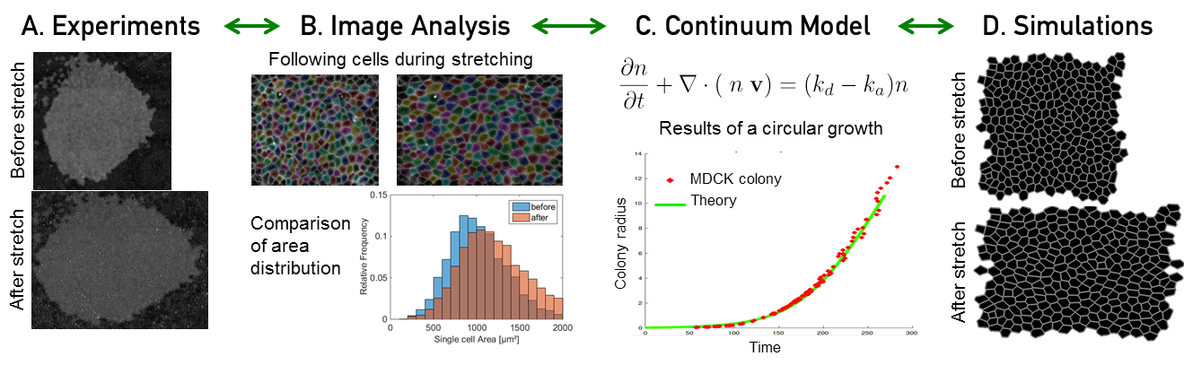
Quantitative analysis and modeling of interaction of epithelial tissues and the environment. (A) An example of experiments for stretching a colony of MDCK cells. (B) Analysis of experimental images with well-established methods to follow individual cells and quantify different properties of cells such as distribution of cell area as plotted here. (C) Continuum models developed to describe the dynamics of the spatial profile of cell density (n) with cell velocity (v) and active terms (k). The results fit very well with the colony growth experiments. (D) Simulation of growth and stretching of tissues at the level of individual cells (vertex model). (A-D) own unpublished data.
Background: A number of studies have highlighted the significance and necessity of quantifications and modeling in biological problems2. Theoretical analysis can help to study living organisms and address the contribution of different mechanisms influencing their morphogenesis48,49. The different models build on standard concepts from different disciplines of physics such as hydrodynamics, elasticity and statistical physics to describe the growth and development of tissues at different time and length scales50. Biological tissues can be seen as active viscoelastic materials. Hence, their elastic and plastic response to deformation, as well as their activity resulting from cell division and apoptosis events, can be modelled by continuum approaches51,52. To obtain detailed information on the force distribution on the level of a cell, these approaches can be complemented by simulations based on vertex or Voronoi models53,54. We recently combined these approaches to study the growth of 2D epithelial tissues and the response to uniaxial stress. Aim: Building on our experience in modeling tissues in 2D, we develop a 3D physical description which will improve our understanding of the cooperation between biochemical signals and mechanical events in the control of the structure, stability and viscoelastic properties of the spheroid. Furthermore, we will investigate the interaction of spheroids with the surrounding planar supports or tissues. We develop a hierarchy of models based on fundamental physical principles taking into account cell mechanics, intercellular interactions and the interactions with the ECM. Accounting for cell division and apoptosis enables us to predict global dynamics during spheroid formation, the transmigration of cells from and into the spheroid, as well as the response to local and macroscopic deformations. Calibrated by experimental data, our theoretical and simulation approaches is used to predict novel features of the system. Approach: This project implements physical and mathematical methods to develop general models describing the time development of epithelial morphology and the interaction of spheroids with external structures. The starting point adapts the known models for epithelial growth, which are usually conceived in planar geometry, and accounting for the change in topology by modeling the closure mechanism necessary to construct 3D objects. Furthermore, we exploit the experimental systems presented in A1-A3 to test and improve models of different complexity - from a simple aggregate of identical cells to spheroids containing different cell types and encapsulated by a basement membrane. By developing sophisticated image analysis protocols, we help to elucidate the role of adhesion and cytoskeletal elements (A1) and the elasticity of the surroundings (A2, A3) on the growth patterns. These data help to build predictive continuum models and simulations, which can then be used to study the dynamics of the system in the conditions that are difficult to explore with other methods, including the response to stress, which is addressed both in 3D (A1-A3) and 2D (A3, B2, B3, D2). Here, a key element is introducing models that can account for the layered structures. As a result, we identify concepts and approaches that can be used in the broader context of the consortium.