Consequences of disease-causing cytoskeletal mutations on epidermal tissue stability
Lead supervisor: R. Leube, Co-supervisor: R. Merkel; Junior supervisor: N. Schwarz Uniklinik RWTH Aachen, Institute of Molecular and Cellular Anatomy (MOCA)
Hypothesis: Blister formation and hyperkeratosis are alternative epidermal mechanoresponses.
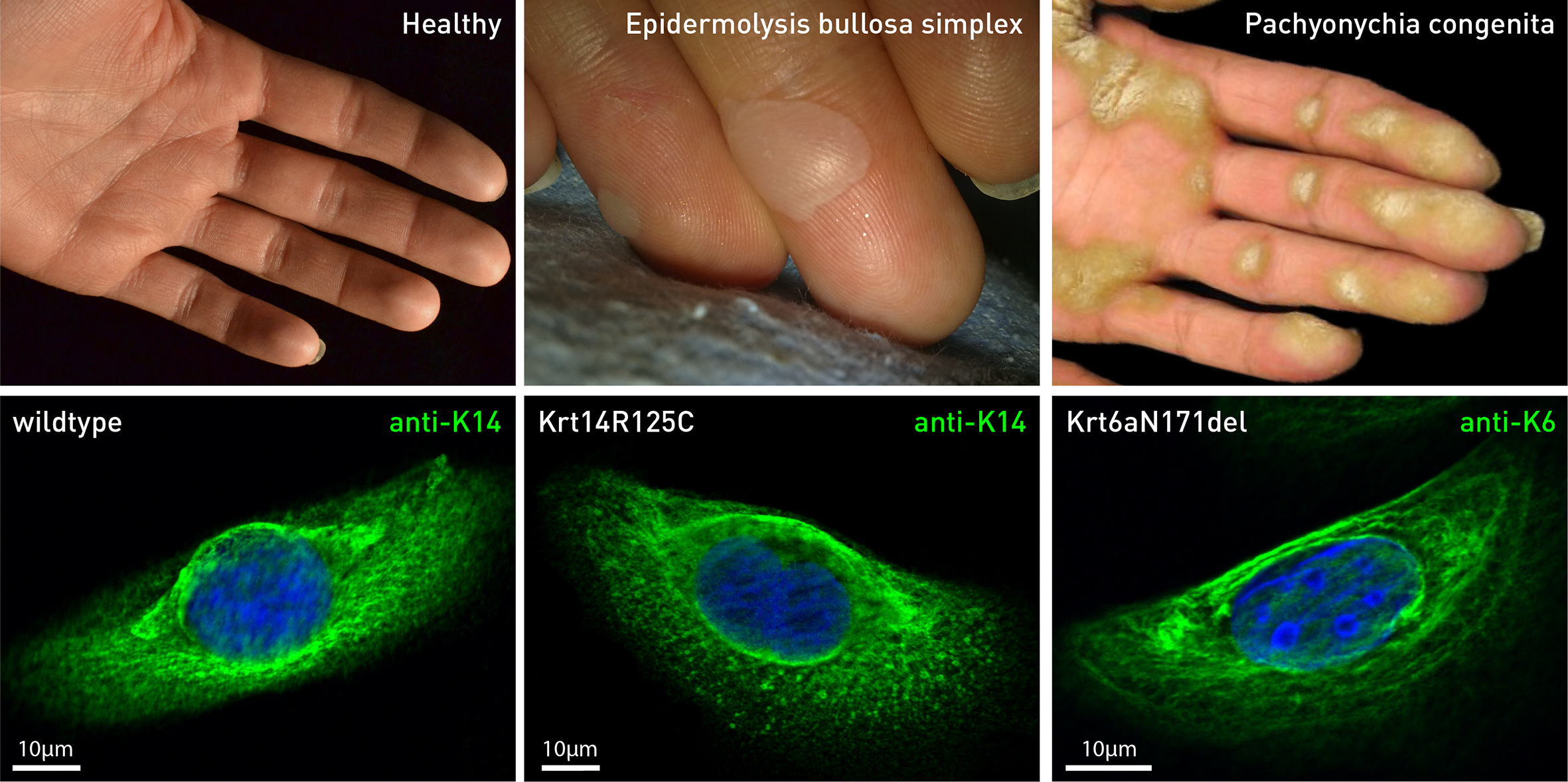
Blister formation and hyperkeratosis in keratinopathies (top; modified images from www) and keratin network morphology in corresponding cultured keratino-cytes (unpublished). Note that keratin networks are detectable by immunofluorescence microscopy with only slight alterations presenting cytoplasmic granules in a keratinocyte derived from an epidermolysis bullosa simplex patient producing Krt14R125C mutant polypeptides and thickened filaments in a keratinocyte derived from a pachyonychia congenita patient producing Krt6aN171del mutant polypeptides.
Background: Keratinopathies are a large group of rare autosomal dominant genodermatoses that are caused by keratin gene mutations. They usually manifest in regions of increased mechanical stress leading to two fundamentally different reactions, which also occur in the skin of healthy humans upon acute and chronic mechanical stress: Blister formation: Compromised keratin network stability results in network disruption and granule formation upon increased mechanical stress eventually leading to cytolysis65. This phenotype is found in epidermolysis bullosa simplex (EBS), which is caused by mutations in keratins K5 and K14. Blister formation is restricted to the K5/K14-rich basal cell layer, which is characterized by a unique mechanophysical environment positioned at the interface between suprabasal cells and the basement membrane. Hyperproliferation and increased cornification: Mutant keratin polypeptides induce an oxidative stress response that is amplified by additional mechanical stress66. Cells react with increased proliferation resulting in elevated cornification. Pachyonychia congenita (PC) is caused by mutations in keratins K6 and K16, which are typically present in the palmoplantar glabrous skin but absent in interfollicular skin. Hyperkeratotic lesions of the palm and foot sole are observed in PC. Our previous work focused on keratin network organization and dynamics in single epithelial cells60,67-70. Imaging routines and analysis tools were developed to quantitatively measure keratin network motility and turnover at subcellular resolution (S. Lehmann, A. Pora and 68,70). Together with FZJ and IWE1, tools were successfully developed and applied to measure cytoplasmic viscoelasticity and overall cellular stiffness60,69. In addition, methods are available to globally or locally modify cytoskeletal networks (pharmacological and genetic manipulation, cell stretcher, magnetic tweezers, laser nanosurgery). Aims: We aim to (i) detect and measure perturbations in keratin network organization and mechanical tissue stability in confluent monolayers and epidermal equivalents derived from EBS and PC patients and (ii) study the basis of compromised mechanical resilience and overshooting mechanoresponse in these prototypic keratinopathies. Approach: Patient-derived confluent monolayers and epidermal equivalents are employed: to examine the effects of keratin mutations on keratin network organization and dynamics. We genetically engineer patient-derived keratinocytes to express fluorescent reporters. To study network dynamics at high 3D resolution, we adapt live imaging chambers for light sheet microscopy (with FZJ). to study local alterations in mechanical properties using the tools and approaches described in B1. This includes traction force microscopy for measuring forces that enter the connective tissue compartment (FZJ), magnetic tweezers for local probing within the 3D constructs (with IWE1), and atomic force microscopy for probing from the surface (FZJ). to investigate altered mechanoresponses using mechanical stressors that are available in B1 (cell stretcher, laser-induced local wounding). Differences in mechanoresponse are tested microscopically and biochemically.