Regulation of the epithelial-endothelial signaling interface by shear stress and substrate stiffness
Lead supervisor: A. Ludwig, Co-supervisor: U. Schnakenberg, Junior supervisors: A. Babendreyer and S. Singh Uniklinik RWTH Aachen, Institute of Pharmacology and Toxicology, Division of Pharmacology in Inflammation
Hypothesis: Endothelial shear stress and ECM stiffness affect signaling at the alveolar epithelial-endothelial interface to support the epithelial barrier.
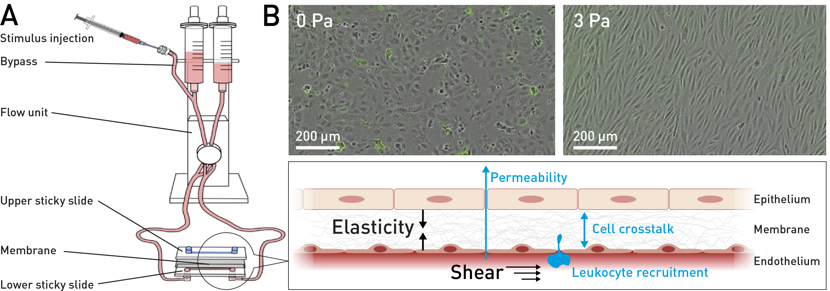
Scheme depicting key aspects of project C1. (A) Established setup for co-culture of endothelial and epithelial cells. (B) The effect of shear and substrate elasticity on permeability, cell crosstalk and leukocyte recruitment will be studied. Flow protects endothelial cells from apoptosis (green cells; unpublished).
Background: The alveolar interface in the lung is formed by the epithelium, the basement membrane and the endothelium. The structural integrity of this interface is crucial for both proper gas exchange and protection against pathogens. Epithelial-endothelial barrier dysfunction clearly worsens the prognosis for patients with inflammatory or fibrotic lung diseases. The epithelial-endothelial interface constantly encounters mchanical forces. This includes endothelial shear stress by blood flow under physiological conditions.75 Moreover, the extracellular matrix between the endothelial and epithelial cell layer is remarkably soft.76 This implies that functions of the epithelial-endothelial interface are critically dependent on flow conditions and tissue softness. It is well understood that pathogens stimulate epithelial cells to produce inflammatory mediators that, in turn, stimulate endothelial cells leading to increased vascular permeability and leukocyte recruitment.77 From previous studies we know that physiological flow conditions can downregulate inflammatory pathways in endothelial cells by preventing the induction of endothelin, adhesion molecules and chemokines,78 by upregulating protective pathways via the transcription factor KLF2 and by regulating ADAM proteases that are known to act on the alveolar interface by cleaving inflammatory effector molecules. Aims: This project aims to decipher signaling events at the epithelial-endothelial alveolar interface in response to flow conditions and substrate elasticity. This will help to understand how the mechanobiological environment contributes to the pathogenesis of inflammatory lung diseases. We examine the contribution of physiological shear stress on endothelial cells and soft substrate to the formation of the tight epithelial barrier. Conversely, we propose that pathologically low shear stress and stiff substrate lead to the generation of mediators that provoke inflammatory endothelial-epithelial crosstalk and elicit an inflammatory response with barrier disruption and leukocyte recruitment. Specifically, we will address KLF2-induced transcriptional responses and the relay function of ADAM proteases. Approach: An established flow chamber system (pump and µ-slides from ibidi) is used to apply physiological (3 Pa) and pathological (0.05 Pa) shear stress to cultured lung microvascular endothelial cells. We have further developed the system by mounting a "sticky ibidi slide" onto the upper side and another onto the lower side of a porous polycarbonate or polyimide membrane (see scheme in the above figure). In this way, the two slides generate two channels separated by the membrane. The channels can be perfused to exert shear stress on cells grown on the membranes. We will now use static and flow systems to combine the culture of epithelial cells on the top of the porous membrane with that of endothelial cells on the lower side (see part B of above figure). Human microvascular lung endothelial cells, lung type 2 epithelial cell lines or primary lung epithelial cells from commercial suppliers are routinely cultured in our laboratory. In search of other more physiological and protective conditions that synergize with flow conditions, we will also include electrospun polycaprolactone or ECM nanofibers. These fibers are produced as membranes with defined elasticity, thickness and mesh size by S. Singh (DWI). By coupling with PEG (with DWI) the elasticity of the membranes are reduced to the kPa range, which best resembles the physiological situation of endothelial cells in their vascular bed.79 These nanofiber membranes are mounted between two sticky slides.