Mechanobiological challenges related to hydrogel-based bioprinting technology for manufacturing novel 3D cell culture models
Lead supervisor: H. Fischer, Co-supervisor: W. Wagner Uniklinik RWTH Aachen, Department of Dental Materials and Biomaterials Research (ZWBF)
Hypothesis: 3D bioprinting techniques affect cellular proliferation and differentiation potential.
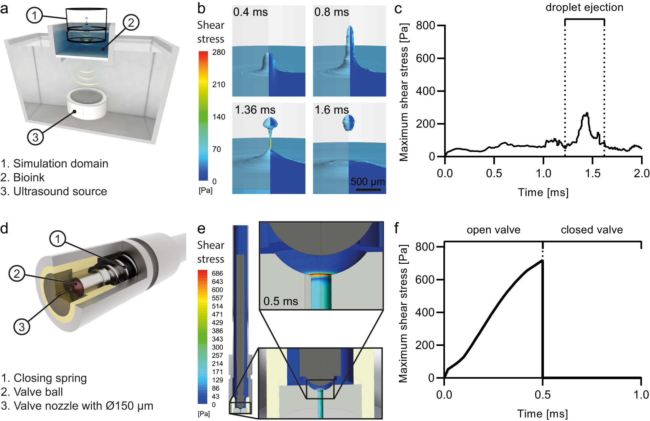
Key aspects of project D2. Shear stress and hydrostatic pressure occur during bioprinting. Such stresses can be harmful to the cells that are embedded in the the hydrogels (bioink). The maximum level of shear stress varies in different 3D bioprinting techniques. (a-c): Acoustic bioprinting technique using acousting droplet ejection principle. (d-f): Inkjet bioprinting technique using microvalves X1.
Background: Hydrogel-based bioprinting technology has gained increasing attention in the field of tissue engineering as it enables the manufacturing of 3D tissue constructs with a defined arrangement of different cell types and materials97,98. This spatial control of distinct regions in the microenvironment has the potential to better mimic the native heterogeneities of native tissues, thereby enhancing the functionality, size, and longlevity of artificial tissues. Besides the use of bioprinted constructs as tissue replacements they are of particular interest for producing advanced 3D in vitro tissue models for analyzing of context-dependent pathophysiology99. We have already successfully bioprinted vascularized tissues, mimetic vessel substitutes, and biofunctional in vitro tumor models100, X2, X3. Despite the encouraging results of the new technology, much has not yet been clarified about cellular responses to the printing process itself. In the first three years of the MEƎT, we were able to show that the maximum level of shear stress strongly varies in different 3D bioprinting techniques X1. Aims: We intend to systematically study the mechanobiological effects of different bioprinting techniques on the proliferation and differentiation of epithelial cells used to prepare the 3D culture models of MEƎT. The role of the dispensing technique and the hydrogel properties, such as viscosity and relaxation behavior, are investigated. Considering the expected insights, the improved potential of 3D bioprinting to realize biomimetic 3D in vitro models are explored. Approach: We use our different custom-made 3D bioprinting platforms (inkjet bioprinting, extrusion bioprinting, and acoustic bioprinting) and our self-constructed bioreactors to investigate cellular responses to the printing-related shear stress and hydrostatic pressure (mechanical signals). We compare different bioprinting techniques, different process parameters, use a variety of natural and synthetic hydrogels and hydrogel blends, and test the effects of cell density. The shear stress and hydrostatic pressure that occur during the printing process are investigated by finite element fluid dynamic models. The mechanical signals are then be correlated with the observed cellular responses.