3D mapping of epidermal tissue mechanics during growth and upon wounding
Lead supervisor: R. Merkel, Co-supervisor: R. Leube Forschungszentrum Jülich, Institute of Biological Information Processing (IBI) 2: Mechanobiology
Hypothesis: Uniaxial, cyclic strain of epidermal equivalents leads to cellular adaptation and, as a consequence, to anisotropic tissue architecture and properties.
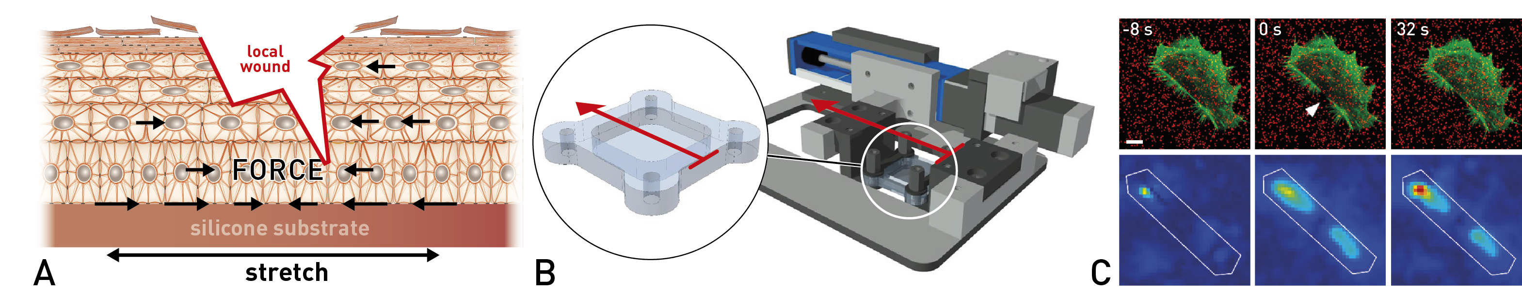
Key features of project B1 (A) Schematic representation of approaches. (B) Cell stretcher. (C) Combination of laser nanosurgery and traction force microscopy (unpublished and 61).
Background: Skin is exposed to intense mechanical stress that is decisive for its development, architecture, and healing. While some basic mechanobiological principles of the dermis have already been established 57, the epidermis is much less thoroughly understood 58. The mechanobiology of epidermal keratinocytes has been mostly studied in subconfluent and confluent monolayers. Our lab has contributed to this by measuring the mechanical properties 59,60and locomotion kinematics 26 of keratinocytes. Moreover, we developed a cell-stretching device for the application of uniaxial cyclic strain 61. While we observed some mechanosensory responses in single keratinocytes, a much stronger reorientation of actin fibers was noted in confluent monolayers. These effects were dependent on adherens junctions together with vinculin and α-catenin as mechanosensors 61. A limitation of these observations is that cell-matrix contacts play a much larger role in single cells and monolayers than in the multilayered epidermis. This was circumvented by other researchers in experiments on intact skin documenting multiple stress-induced effects such as load-dependent cell division axes 62, 63. In a study combining imaging on intact skin, mechanical measurements on keratinocyte monolayers and mathematical modeling of the interplay of cortical tension, cell division and cell-cell adhesion was shown to be decisive for the layered organization of the epidermis 63. However, as all experiments on skin relied on cell-generated forces under static conditions, the adaptation of epidermis to defined external mechanical signals could not be studied. Aims: We aim to discover how uniaxial cyclic strain influences epidermal organization, passive and active mechanical tissue properties, and wound healing, e.g. via anisotropic cell properties and division. The final goal is to connect observable tissue parameters (organization, stiffness, healing, and prestress due to active cell contraction) to cellular processes to finally understand the mechanical adaptation of the epidermis. Approach: Initial experiments are performed on confluent monolayer cultures to measure cellular processes and forces during healing in 2D. Traction force microscopy 6, 43, 44is combined with cell stretching and laser nanosurgery to assess the influence of strain on healing of model wounds in these monolayer systems. For subsequent experiments, simplified epidermal equivalents (SEE) are grown in elastomeric chambers. In these, SEE are exposed to mechanical strain of physiological intensity for extended periods of time. The mechanically conditioned constructs are analyzed by live-cell microscopy and immunofluorescence microscopy. Finally, laser nanosurgery creating microscopic wounds of defined geometry and size at the surface and within epidermal equivalents and subsequent microscopic monitoring is conducted to study layer-specific wound healing processes at high spatial and temporal resolution. Analysis of ensuing tissue relaxation will serve to map local tension patterns and their expected anisotropy.