Mechanobiology of differentiation and invasion in 3D cell assemblies

This project area aims to contribute to the understanding of the cross-talk between local mechanical cues and adaptation/differentiation in multilayer epithelia. The selected paradigm is the epidermis, which is by far the most extensive epithelial tissue surrounding the entire outer body surface. It is characterized by the arrangement of tightly-coupled keratinocytes in layer-specific configurations, each with unique differentiation features and mechanobiological properties. To work toward elucidation of mechanical cross-talk mechanisms, novel tools have been and still need to be developed to enable layer-specific analyses. Another focus of this project area is the dysregulation of this balance in human diseases, which are often associated with perturbed neuronal perception resulting in pain and itch. The underlying pathomechanisms of mechanosensation and mechanotransduction are not known.
Consequences of keratin mutations on epidermal tissue mechanics and mechanoresponses
Institute of Molecular and Cellular Anatomy (MOCA), Uniklinik RWTH Aachen
Rudolf E. Leube
Principal Investigator
Nicole Schwarz
Junior Supervisor
Elena Honscheid
Associated Doctoral Researcher
Keratins as organizers of cytoskeletal space
Sungjun Yoon
Associated Doctoral Researcher
Combining image restoration and traction force microscopy to study extracellular matrix-dependent keratin filament network plasticity
Kyeongmin Kim
Doctoral Researcher
Consequences of keratin mutations on epidermal tissue mechanics and mechanoresponses
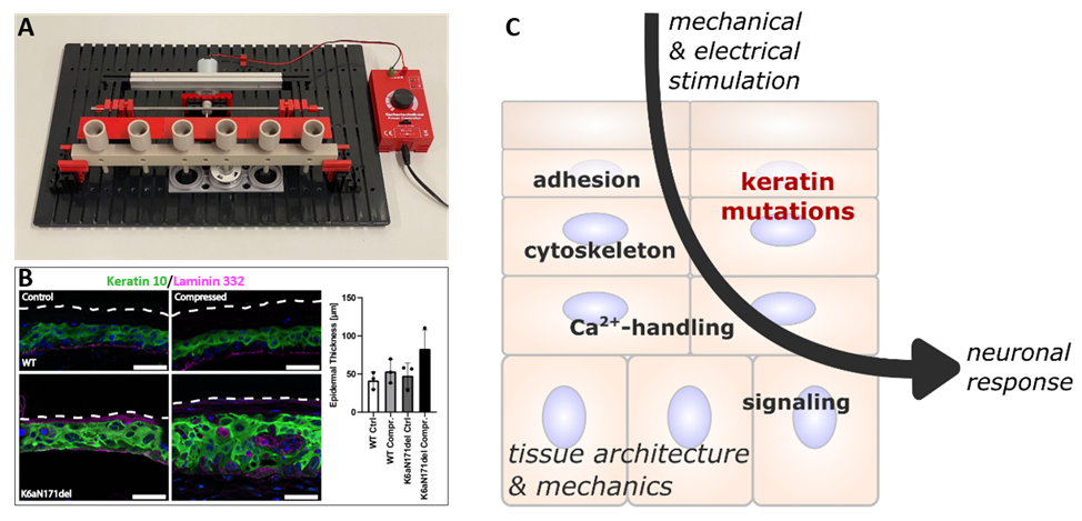
Project overview. (A) shows the high throughput compression devise designed and constructed by the team of Horst Fisher (project area D). (B) highlights the altered response in Pachyonychia congenita (PC)-derived epidermal equivalents. The fluorescence micrographs show sections of wild-type (WT)- and PC-derived (K6aN171del) skin models that were subjected to cyclic compression (47 mbar, 150 mHz, 1h/day, 5 days) or not. The epidermal equivalents were immunostained 4 days after mechanical stress with antibodies against laminin 332 (magenta) to delineate the basement membrane and against keratin 10 (green) to indicate vital suprabasal epidermal layers. Nuclei are stained with Hoechst 33258 (blue). The outermost border of the stratum corneum is demarcated by a dashed line. Scale bars,  50 µm. The histogram at right quantifies the mechanical stress-induced enlargement of the epidermal compartment (including the stratum corneum) in the PC-derived model (mean ± SD). (C) summarizes aspects that are addressed experimentally in the project.