Mechanobiology of differentiation and invasion in 3D cell assemblies

This project area aims to contribute to the understanding of the cross-talk between local mechanical cues and adaptation/differentiation in multilayer epithelia. The selected paradigm is the epidermis, which is by far the most extensive epithelial tissue surrounding the entire outer body surface. It is characterized by the arrangement of tightly-coupled keratinocytes in layer-specific configurations, each with unique differentiation features and mechanobiological properties. To work toward elucidation of mechanical cross-talk mechanisms, novel tools have been and still need to be developed to enable layer-specific analyses. Another focus of this project area is the dysregulation of this balance in human diseases, which are often associated with perturbed neuronal perception resulting in pain and itch. The underlying pathomechanisms of mechanosensation and mechanotransduction are not known.
3D-mapping of epidermal tissue mechanobiology during growth and upon wounding
Helmholtz-Institute for Biomedical Engineering, Division of Stem Cell Biology
Rudolf Merkel
Principal Investigator
Hajaani Manoharan
Doctoral Researcher
Epidermal cell layers under strain: Analysis and manipulation of mechanoresponse processes in healthy and diseased biomimetic tissue systems
Pauline Eichstedt
Doctoral Researcher
Exchange kinetics of desmosomal proteins under the influence of mechanical stretch
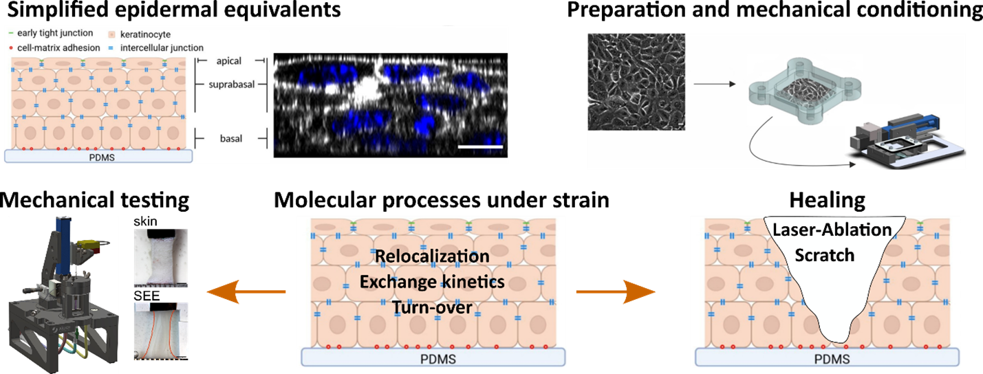
Project overview. Simplified epidermal equivalents are prepared from primary and immortalized healthy keratinocytes or mutated keratinocyte cell lines. They are preconditioned by cyclic mechanical stretching in a self-made device. Subsequently, mechanical, and molecular properties are quantified under external strain and during healing of model wounds. Parallel experiments on skin ensure physiological relevance.